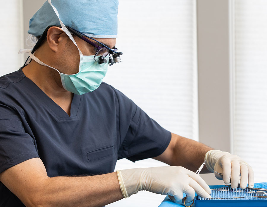
Why Reconstructive Surgery
The need to undergo oculofacial reconstructive surgery is often dictated beyond one’s control—frequently due to trauma, cancer, or the way one was born.
Our team has extensive experience in treating patients who need reconstructive procedures for eyelid malpositions, facial skin cancer, orbital/facial trauma, lacrimal (tearing) problems, Thyroid Eye Disease, and scarring. The situation surrounding a reconstructive procedure can be challenging, and we accompany you through the process to ensure that your needs are met, and that you achieve the best outcome possible. Undergoing a oculofacial reconstructive procedure is often not a choice, but it can be a successful experience.
Skin Cancer
Eyelid & Facial Reconstruction for Skin Cancer
The eyelid skin is the thinnest and most sensitive skin on your body.
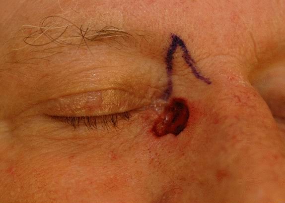
As a result, this is often the first area on your face to show change from sun damage and aging. Unfortunately, sun damage and other environmental toxins not only cause the skin to age but can cause serious damage. Skin cancer of the eyelids is relatively common and several types exist. The presence of a nodule or lesion on the eyelid that grows, bleed or ulcerates should be evaluated. This involves examination and sometimes a biopsy.
Basal Cell Carcinoma
Basal cell tumors represent the ninety percent of eyelid tumors. These skin cancers grow slowly over months and years. They most often appear as a pearly nodule that eventually starts to break down and ulcerate. Despite being a cancer, these tumors don’t spread to distant areas but rather just continue to grow and infiltrate the surrounding tissue. They typically can be cured by simple excision followed by reconstruction of the defect left behind after the tumor removal.
Squamous Cell Carcinoma and Melanoma
These types of tumors occur much less common but are more aggressive and require more involved care to ensure complete treatment. Again, primary treatment involves removing the tumor, but care must also be taken to ensure the tumor has not spread anywhere, causing larger health problems. Your surgeon will help coordinate this as part of your treatment depending on the size and circumstances of the tumor at presentation.
Tumor Excision and Eyelid Reconstruction
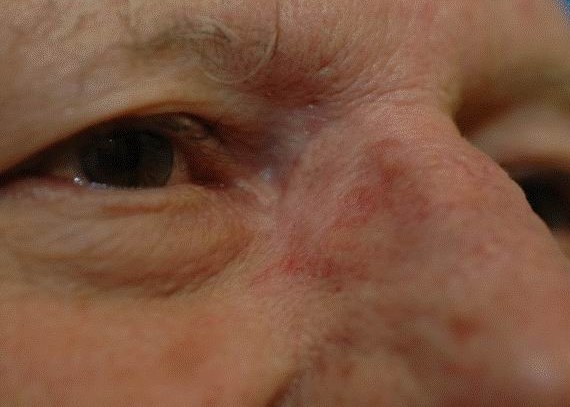
Skin cancer needs to be removed surgically by a skilled individual who can not only remove the tumor but reconstruct the eyelid or area where the tumor was removed. Sometimes surgeon will do this themselves at a surgical facility with an on site pathologist who can immediately examine the specimen to ensure the whole tumor was removed. Other times, the help of a dermatologic surgeon specializing in Mohs surgical excision will be utilized. This procedure is completed into two steps, the first in the dermatologist’s office with immediate examination of the tumor to ensure its complete removal followed by the reconstructive surgery by your surgeon.
Eyelid reconstruction may be undertaken in a variety of situations. Defects in the eyelid may arise form a variety of situations, but most commonly after trauma or tumor excision. Simple superficial defects in the eyelid may occur after minor trauma or removal of small growths. Many of these require nothing more than local wound care and will heal on their own in a week to 10 days. Some simple superficial defects may require a few sutures with the same local wound care.
In some instances, such as after traumatic injuries or removal of larger growths or skin cancers, larger defects may extend through the entire lid. Many of these can be sutured together directly, but many others may require more complex reconstructions. In many of these more complex cases, the surgeon will need to use transfer of adjacent tissues (what we call ”flaps”), or transfer of skin from other parts of the eyelid face or body (what we call “skin grafts”) to complete the reconstruction. Some of these more complex reconstructions may require more than one operation to complete (what we call “staged reconstruction”).
Thyoid Eye Disease
Also known as Thyroid Orbitopathy or Graves’ Disease
Thyroid eye disease is an autoimmune inflammatory disorder of uncertain etiology that affects the tissues of the orbit (i.e. eyelids, eye muscles and other soft tissues surrounding the eyes). Patients with this disorder often have an associated thyroid abnormality which may manifest either before, during or after the orbital signs and symptoms. However, a small percentage of patients may have eyelid and orbital manifestations of the disorder without developing a thyroid abnormality.
Disease Characteristics
Thyroid orbitopathy can develop and affect patients with varying degrees of severity. The disease can begin suddenly and progress rapidly over days to weeks or start insidiously and progress gradually over a long period of time. The majority of patients have mild inflammation, the most common signs and symptoms of which are retraction of the upper and/or lower eyelids and bulging of the eyes (also known as proptosis). In cases of moderate inflammation, patients may also have varying degrees of double vision and eyelid swelling as well as visible redness of the lids and eyes. Other common signs and symptoms include, redness, dryness, excessive mucous and tear production, and pain/pressure behind the eyes. A small percentage of patients with thyroid eye disease develop severe inflammation with massive enlargement of the eye muscles which can result in compression of the optic nerve and, and possible, permanent vision loss. In most cases, however, the inflammatory process is self-limited and runs a course lasting 6 months to a couple years before subsiding. After the inflammatory phase of the disease subsides, scarring of eyelid and orbital tissues may result in the persistence of eyelid retraction, proptosis and double vision.
Evaluation and Management
It is important to be evaluated by an ophthalmologist to assess the ophthalmic manifestations of the disease as well as by an endocrinologist to manage concurrent thyroid abnormalities. Patients with mild Thyroid Eye Disease are usually evaluated on a 3 to 4 month interval basis to monitor progression of the disease and managed with measures to reduce ocular symptoms. Patients with moderate to severe Thyroid Eye Disease may require medical or surgical intervention to reduce inflammation or improve vision. Medical treatment starts with more conservative measures of lubricating eye drops, cool compresses, and non-steroidal anti-inflammatory medicine such as Ibuprofen or Naproxen. One of the most important non-invasive, though challenging, interventions in combating disease severity is smoking cessation. Cigarette smoking has been associated with development of worsening thyroid orbitopathy and increased risk of vision loss, and the importance of therefore cessation of smoking cannot be overstated.
As disease severity intensifies, IV or oral steroids (prednisone and related medicines) may be required. There are a variety of immune modulating drugs that are now available to mitigate the inflammation, but most are still considered experimental or second line agents.
Once the inflammatory phase of the disease has subsided, surgical rehabilitation can be initiated. Patients with eyelid abnormalities, double-vision or proptosis may be eligible for surgical correction to improve their function and appearance, usually performed in a staged fashion. Orbital decompression [bone and/or fat] will first be considered to allow the eyes to settle back in the eye socket and decrease bulginess. Once that has been achieved, eye muscle surgery will be performed in patients that have bothersome double vision (diplopia). Eyelid retraction repair will then be performed to improve eyelid position to allow better eyelid closure. As the disease process will cause enlargement of the eyelid fat and skin, blepharoplasty is often performed to improve cosmesis.
Eyelid Malposition—Entropion & Ectropion
Entropion
Entropion is a condition in which the eyelid is rolled inward toward the eye. It can occur as a result of advancing age and weakening of certain eyelid muscles. Entropion may also occur as a result of trauma, scarring, or previous surgeries. Entropion may also occur in children.
A turned in eyelid rubs against the eye, making it red, irritated, painful, and sensitive to light and wind. If it is not treated the condition can lead to excessive tearing, mucous discharge and scratching or scarring of the cornea. A chronically turned in eyelid can result in acute sensitivity to light and may lead to eye infections, corneal abrasions, or corneal ulcers. If entropion exists, it is important to surgically repair the condition before permanent damage to the eye occurs.
Surgical Repairs
There are a number of surgical techniques for successfully treating entropion, depending on the cause and presenting anatomic problem. The most common surgical treatment involves tightening of the eyelid attachment to the eye socket, while reinforcing the muscle attachments on the inside of the eyelid to restore the lid to its normal position. The surgery to repair entropion is usually performed as an outpatient procedure under local anesthesia or with IV sedation. Patients recovery quickly using an antibiotic ointment for about one week after surgery. Most patients experience immediate resolution of the problem following surgery.
In certain situations, a non-incisional entropion repair, using only sutures, may be performed as an in-office procedure under local anesthesia. This procedure, sometimes referred to as the “Quickert” procedure requires several strategically placed sutures which externally rotate the eyelid. This procedure is an excellent treatment for patients who are not suitable for surgery, or until more definitive surgery can be performed.
Ectropion
Ectropion is an eyelid malposition indicating the lower eyelid is “rolled out” away from the eye, or is sagging away from the eye and the wet, inner surface of the eye (conjunctiva) may be exposed and visible. Normally, the upper and lower eyelids oppose each other, lubricating and protecting the eye. If the edge of one eyelid turns outward, the two eyelids cannot meet properly and tears are not spread evenly over the eye, and the sagging lower eyelid may leave the eye exposed and dry. If the ectropion is not treated, the condition can lead to chronic tearing, eye irritation, redness, pain, a gritty feeling, crusting of the eyelid, mucous discharge, and breakdown of the cornea due to exposure.
Orbit (Eyesocket) & the Tear Duct System
Orbital Trauma
In the United States, injuries to the eyes and eye socket are unfortunately common place. Eyelid laceration, tear drain injuries, bleeding within and bruising around the eye, and fractures of the bones of the eye socket can all occur. These injuries often occur from sporting activities like baseball, soccer or football. In addition car accidents, fist fights, and dog bites are other common causes. Trauma to the orbit and eye area most often results in bruising, eyelid lacerations, and fractures of the orbital bone Since the bones of the eye socket done move like your arm or leg, they don’t all need to be fixed. Fractures causing double vision or large fractures into the sinus should typically be repaired in a timely fashion. Sometimes this is not possible but late repairs, despite being more difficult, can still be done.
Sometimes trauma is severe enough to irreparably injure the eye. Along with diseased eyes that are blind, painful and disfiguring, the removal of an eye (enucleation) is indicated when the eye can not be salvaged. Reconstruction of the eye socket, followed by fabrication of a prosthetic eye (performed by an ocularist), typically leads to a very pleasing result. Regular examinations are required as occasionally further reconstruction may be needed to retain proper fit of an eye prosthesis.
Orbital Tumors & Infections
Orbital infection, or Orbital cellulitis, is an aggressive sight, and even life, threatening process. Usually arising secondarily from an adjacent sinus infection, this situation must be treated swiftly and aggressively with antibiotics and often surgery. Close follow up and monitoring is required to treat these infections, but is typically successful. Of note, not all infections occur as an extension of sinusitis. This can also occur after trauma to the eyelid, eye or orbit as well as in patients whose immune system is challenged.
In both children and adults, a variety of tumors can occur in the eye socket. Some grow slowly, and go unnoticed while others can grow rapidly; impairing vision and causing even greater problems. CT scans and MRI’s are the best method for detecting and differentiating these lesions prior to having surgery. Once the location is identified, along with the general characteristics of the lesion, a treatment plan can be created. In some instances your orbital surgeon can treat these tumors on their own, often as an outpatient. On the other hand, aggressive tumors may require the help of other surgical specialist and in patient hospital treatment.
Tear Duct System—The Tearing Patient
The tear film on the surface of the eye is a critical component of maintaining vision. Tears nourish and lubricate the surface of the eye as well as wash away debris. A smooth, balanced tear film (consisting of water, oil and mucus) also allows light to enter the eye in an optimal fashion. If there is a disturbance of the tear film, patients will often experience tearing, burning, irritation and most importantly blurred vision, which often fluctuates. Patients who experience tearing either have a problem with ocular surface lubrication and tear production, or tear drainage.
Increased Tear Production and Dry Eyes
The eye has two sets of structures that produce tears. Smaller tear glands help maintain a baseline level of moisture on the surface of the eye. Unfortunately, inflammatory conditions like rheumatoid arthritis, Sjogrens disease as well as aging and menopause lead to decreased tear production. As tear production diminishes, the surface of the eye starts to dry out. Further, inflammation of the oil glands along the edge of the eyelid, common in patients with roseacea, also causes early breakdown and evaporation of the tear film. The brain senses the eye is both dry and irritated and in turn signals the main tear gland to flush the eye. As a result, the dry eye paradoxically tears and becomes watery. Patients with dry eyes note intermittent tearing of the eyes during activities like reading, driving, watching TV, using a computer or going outside on a windy day. These all cause the eye to dry out because the eye blinks less during these activities. The treatment for dry eyes includes 1) replacing tears with artificial lubricants which can be bought over the counter, 2) medications like Restasis that decrease inflammation in tear glands and encourages natural tear production to resume and finally 3) plugging of the tear drain. Other causes of increased tear production exist like allergies, infections and eyelashes poking the eye. These conditions can often be found during examination.